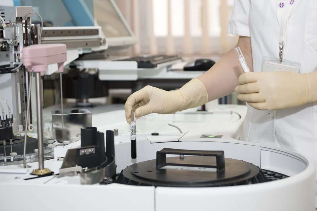
Microbiological Testing is a mandatory requirement to ensure the quality of the packaging material which has a direct impact on the quality of a drug. The goal of the packaging material is to protect the contents against external factors such as humidity, light, oxygen or temperature variations. Microbiological Testing of the packaging material is done to ensure that the product protected against external impacts or not.
The packaging material itself should neither interact with the contents of the packaging nor should it have a negative influence on the contents. Ideally, there will also be no transfer of ingredients from the packaging material to the drug. I will elaborate on how to conduct microbiological testing on empty bottles, droppers, and tubes.
Procedure
The procedure to conduct microbiological testing of the packaging material is as follows.
A For Total Microbial Count
A.1 Method Applied: Pour Plate
A.1.1 Fill each container with peptone and pour the contents in a pre-sterilized flask.
A.1.2 Aseptically takes 1ml of the sample from (A.1.1) in each of four Petri plates.
A.1.3 Add (15 — 20ml) of liquid trypticase soy agar on the sample’s taken in 2 Petri plates for bacterial growth.
A.1.4 Swirl the plate gently, cover, after solidification, invert to remove moisture from the plate and incubate at 30° — 35°C for 3 to 5 days.
A.1.5 Similarly repeat the step (A.1.3 — A.1.4) for sabouraud dextrose agar (SDA) in other two plates for fungal growth and incubate at 20° — 25°C for 5 to 7days.
Evaluation
After completion of incubation, count the colony-forming units taking an average of two Petri plates for each agar medium and record the results.
B For Pathogenic Bacteria
B.1 Staphylococcus aureus and Pseudomonas aeruginosa
B.1.1 Aseptically transfer 10 ml of the sample from (A.1.1) into 90ml trypticase soy broth, disperse and incubate at 30° — 35° for 24 to 48 hours.
B.1.2 If growth is present in (B.1.1) mix gently and streak on:-
i- Mannitol Salt Phenol Red Agar Medium —— Staphylococcus aureus
ii- Cetrimide Agar Medium —– Pseudomonas aeruginosa
B.1.3 Cover, invert and incubate the dishes at 30° — 35°C for 24 to 48 hours.
Evaluation
Examine any resulting growth.
Staphylococcus aureus
- Staphylococcus aureus gives yellow colonies with yellow zones on Mannitol Salt Phenol Red Agar Medium. Then perform a gram staining test.
- Gram Staining Test: It should be positive cocci in the cluster
Confirmation
To confirm the Staphylococcus aureus Biochemical differentiation test (API Staph) is performed.
Pseudomonas aeruginosa
- Pseudomonas aeruginosa generally gives green colonies on Cetrimide Agar Medium. If fluorescence is checked in ultraviolet light, it will be greenish. Then perform a gram staining test.
- Gram Staining test: It should be gram-negative rods.
Confirmation
To confirm Pseudomonas aeruginosa, Oxidase test is performed.
Oxidase test
- Transfer the colony to be tested to an Oxidase Detection Strip using a platinum wire loop.
- Spread the culture on the strip and observe for up to 5 seconds.
A deep blue / violet color indicates a positive reaction.
The presence must be confirmed by API 20 NE (Biochemical differentiation test).
B.2 Salmonella Species
B.2.1 Aseptically add 10ml of the sample from (A.1.1) in 90ml lactose broth, disperse and incubate at 30° — 35°C for 24 to 48 hours.
B.2.2 If growth is present mix gently and pipette 1ml portion into vessels containing 10ml of Selenite Enrichment Broth (double strength)
B.2.3 Subculture onto any of two plates of each medium by streaking on.
- Brilliant Green Agar Medium
- Xylose Lysine Desoxycholate Agar Medium
- Bismuth Sulfite Agar Medium
Cover, invert and incubate the Petri plates at 30° — 35°C for 24 to 48 hours.
Evaluation
1.0 On Brilliant Green Agar Medium Salmonella give small, transparent, colorless or pink to white opaque colonies (frequently surrounded by pink to red zone).
2.0 On XLD Agar Medium, Salmonella gives red colonies that are with or without black centers.
3.0 On Bismuth Sulfite Agar Medium, Salmonella give black or green colonies.
Confirmation
To confirm Salmonella, transfer the suspected colonies with the help of an inoculating wire to a butt slant of Triple Sugar Iron Agar Medium. A first streak on the surface of the slant. Stabb the wire well beneath the surface of the slant. Incubate at 30° — 35°C for 24 to 48 hours.
If the slants become alkaline (red) and butt become acidic (yellow), with or without concomitant blackening of the butt from hydrogen sulfide production, it indicates the presence of genus Salmonella.
Confirm the results by using API 20E (biochemical differentiation test).
B.3 Escherichia coli
Transfer quantity of the contents A.1.1 corresponding to 1gm or 1ml to 100ml of enrichment medium (Enterobacter enrich broth Mossel) and Incubate the (EBM) and remaining lactose broth at 35 — 37°C for 24 to 48 hours.
If the growth is present in EBM mix gently subculture on two plates of Levine EMB Agar and incubate at 35 – 37°C for 24 to 48 hours.
Evaluation
On the Levine EMB Agar, colonies suspected of being Escherichia coli appear greenish metallic sheen in reflected light, dark or even black center in transmitted light. Perform the gram staining it should be gram-negative rods.
Confirmation
The biochemical differentiation test is performed to confirm Escherichia coli (using the API 20 E system).
Requirements
A. Total Microbial Count < 50 CFU
B. Pathogenic Bacteria Acceptance Criteria
Staphylococcus aureus —- No growth
Pseudomonas aeruginosa—- No growth
Salmonella species—- No growth
Escherichia coli—– No growth

