Drinking Water
Drinking water safety is a major concern throughout the world. We all are well aware that 90 percent of diseases are water born. Therefore drinking water should be free from pathogens. In this article, I will explain how to test drinking water in a microbiology laboratory.
Preparation of Apparatus
Thoroughly wash and finally rinsed glass apparatus e.g., Petri dishes pipettes, flasks, graduated cylinders with purified water and sterilized in a dry heat oven at 160ºC for 120 minutes or 170º to 180°C for not less than 60 mins.
Filter units are thoroughly washed with purified water and wrapped loosely with Parchment Paper.
Filter holders, scissors, forceps, with Parchment Paper, and along with wrapped filter units sterilize by autoclaving at 121°C for 30 minutes.
Excessive or prolonged heating will damage the filters. It may be convenient to sterilize all the above equipment and apparatus in a suitable metal container.
Procedure
Total Microbial Count (Pour Plate Method)
Using a sterile pipette, aseptically add 1ml of the sample into two Petri plates (run in duplicate).
Aseptically add 20 – 25ml of melted R2A Agar (at about 45°C) in Petri dishes containing water samples.
Swirl the plate gently, cover after solidification, invert and incubate at 30 – 35°C for 48 – 72 hrs.
NOTE: The test should be conducted under LFC.
Evaluation
After completion of incubation, count the colony-forming units taking an average of two Petri plates for each agar medium and record the results.
Membrane Filtration Method (Alternate Method)
The sterilized filter of porosity (0.45mm) is aseptically placed on the filter base of the filtration assembly while the ration unit is attached to a vacuum pump. The membrane is first rinsed with 100ml of sterile Drinking water and then pour 1ml of sample to be tested (diluted sample can be tested if the count is expected).
Rinsed the filter membrane again with drinking water after filtration of the sample (in membrane filtration technique for drinking water testing quantity can be increased that is instead of 1ml, 50ml or 100ml can be used, if not very high count is expected).
Disassemble the filtration assembly, remove filter aseptically and placed to R2A Agar plate, incubate at 30 – 35°C for 48 – 72 hours.
Evaluation
After completion of incubation, count the colony forming units (the result evaluation of membrane filtration will be the total count obtained from the single filter membrane).
Confirmation
If the number of viable micro-organisms increased the alert limit than First Streak the morphologically identical colonies which are more in count on Tryptic Soy Agar and incubate at 30 to 35°C for 24 hours.
Perform the gram staining.
If morphological identical colonies are gram-negative rods confirm them with biochemical differentiation test (API 20 E & API 20 NE).
If the colonies are gram-positive cocci confirm them with API Staph.
Pathogenic Bacteria Identification
Psudomonas aeruginosa
Add 50ml of water to 25ml Malachite Green Broth (Triple Strength). Thus the final concentration of inoculated broth will always be single strength. Incubation at 35°C ± 1°C for 24 – 48 hours.
From the incubated Malachite Green Broth, subculture onto the one plate of cetrimide agar. Cover, invert and incubate the dishes at 30 – 35°C for 24 – 48 hours.
Examine the resulting growth as following
Pseudomonas aeruginosa generally gives green colonies on Cetrimide Agar Medium. If fluorescence is checked in ultraviolet light, it will be greenish. Perform gram staining, it should be gram-negative rods.
Confirmation
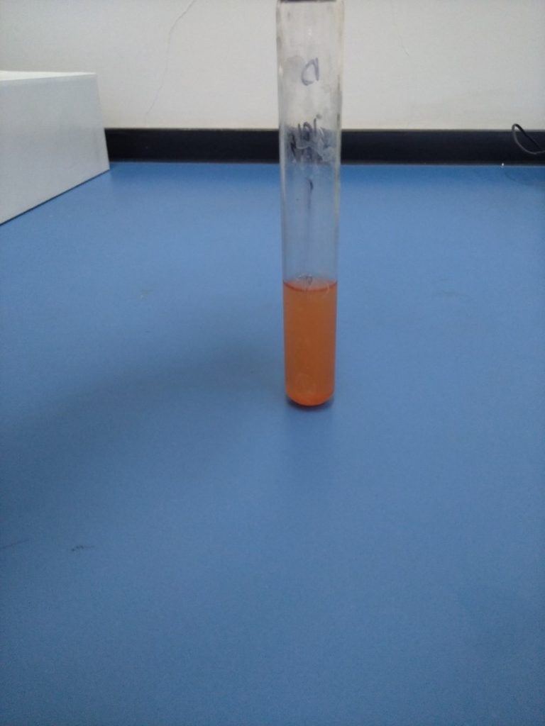
To confirm Pseudomonas aeruginosa Oxidase test is performed.
Transfer the colony to be tested to an Oxidase Detection Strip using a platinum wire loop.
Spread the culture on the strip and observe for up to 5 seconds.
A deep blue / violet color indicates a positive reaction.
The presence must be confirmed by API 20 NE (Biochemical differentiation test).
Salmonella Species
Aseptically add 10ml of the sample in 90ml lactose broth, disperse and incubate at 35 – 37°C for 24 – 48 hours.
If growth is present mix gently and pipette 5ml sample with double strength portions into tubes containing 10ml selenite cystine broth in the ratio 1:1, mix and incubate at 35 – 37°C up to 24 hours.
Subculture on any of two plates of each medium by streaking.
a- Brilliant Green Agar Medium.
b- Xylose Lysine Desoxycholate Agar Medium
c- Bismuth Sulfite Agar Medium
Cover, invert and incubate the Petri plates at 30 – 35°C for 24 – 48 hours.
Evaluation
On Brilliant Green Agar Medium, Salmonella gives small, transparent, colorless or pink to white opaque colonies (frequently surrounded by pink to red zone).
XLD Agar Medium, Salmonella gives red colonies that are with or without black centers.
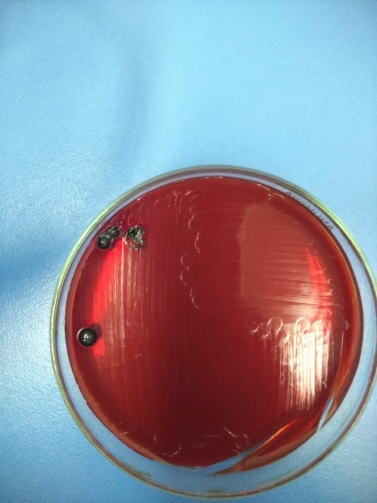
Bismuth Sulfite Agar Medium, Salmonella gives black or green colonies.
Perform gram staining; it should be gram-negative rods.
Confirmation
To confirm Salmonella, transfer the suspected colonies with the help of inoculating wire to a butt slant of Triple Sugar Iron Agar Medium. A first streak on the surface of the slant and then stabbing the wire well beneath the surface; incubate at 30 – 35°C for 24 – 48 hours.
If the slants become alkaline (red) and butt become acidic (Yellow), with or without concomitant blackening of the butt from hydrogen sulfide production, indicating the presence of genus Salmonella.
Coli Form and E. coli
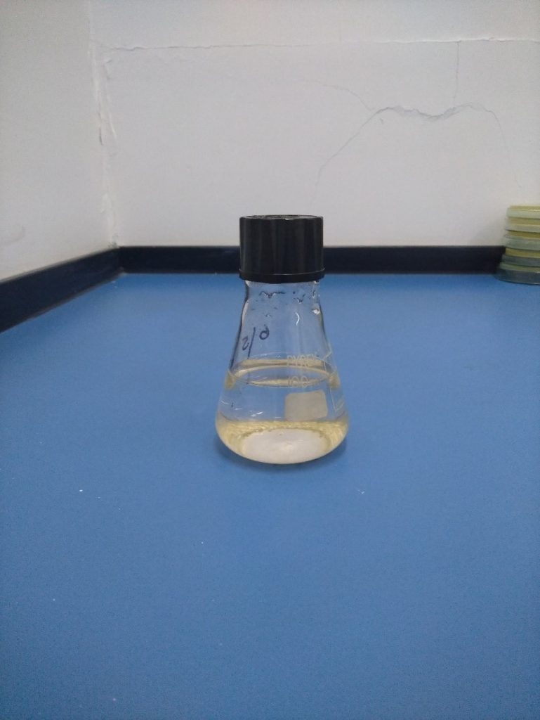
Add 50ml of water sample into a sterile, transparent 100ml tube/flask with a screw cap.
Attention: Glass apparatus is not shelf fluorescence.
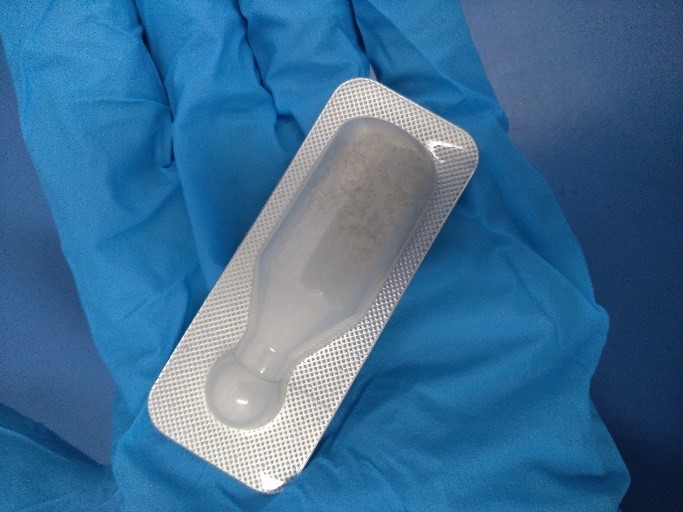
Take one snap pack of media Readycult® Coliforms. shortly tap to ensure the granules are at the bottom. Bend the upper part of the snap pack until at break opens.
Attention: Do not touch the opening to avoid contamination risk
Add the content to the water sample seal the vessel and shake to dissolve the granules completely (broth is clear & yellowish).
Incubate 18 – 24 hours at 35°– 37°C.
Interpretation of Results
| Negative——- No Color Change |
| Total Coli Form—–Color Change to Blue-green (No discoloration with shaking) |
| E. coli——Check blue-green colored vessel for fluorescence by the UV lamp. To confirm the E. coli overlay the broth with 2.5 ml of KOVAC’s reagent (Indole reaction). A red ring confirms presence of E. coli. Light blue fluorescence indicates presence of E. coli. |
Acceptance Criteria
Total Microbial Count = <500 cfu/ml
Coli Form/ E. coli = Absent
Salmonella Species = Absent
Pseudomonas aeruginosa = Absent